 |
Available
at our Lahore Facility
contact us
|
Although the researchers have not clearly established how mycoplasmas are transmitted, they have made some inferences from the data they have collected.
The mycoplasma organism has been found in the blood and body fluids, spinal fluid, bone marrow, urine, and in the lungs, nose and mouth. The mycoplasma is reported to be able to survive for two hours outside the body (Baseman & Tully, 1997) .
Some sort of chemical exposure or immune distress (i.e., auto accident, surgery, cancer) appears to pre-date the onset of illness. Of those with CFS, FMS, GWS, and RA, numerous friends and spouses have the illness, as well as close relatives (Nicolson, Nicolson & Nasralla, 1998; Nicolson, et al., 2000).
When examining those with GWS, it was noted that immediate family members also had similar symptoms (Nicolson, et al., 2000). Several babies have also been known to be born with a similar illness (Nicolson & Nicolson, 1997; Nicolson, Nicolson & Nasralla, 1998). In a more recent study of immediate family members of those with GWS, when the blood was examined, it was found that most family members of GWS patients that were symptomatic with signs and symptoms of CFS also had mycoplasmal infections. Of those diagnosed with GWS, 82% were infected with mycoplasma as was 77.5% of family members with symptoms of CFS. In both groups, the predominate mycoplasma species found was M. fermentans. (Nicolson, et al., 2003). Speculation as to how those with GWS contracted the mycoplasma infection has led to questioning immunization practices of military personnel and/or possible biological exposure during deployment (Nicolson, et al., 2000). Inferences made from studies of immediate family members of those GWS veterans who have mycoplasma infections, suggest that casual transmission is possible.
Therefore, it would appear that mycoplasma is transmitted after deployment in the military, through casual contact with one who is infected with mycoplasma, and both casual and intimate contact and from mother to fetus. This means that the organism may possibly be contracted from vaccines contaminated with mycoplasma or from contamination from biological weopons. It can be passed from person to person through sputum (coughing droplets that contain the organism), saliva, sexual secretions, blood, and urine.
(Created 6/10/05 by Sharon Briggs)
-
Cancer Caused
By Bacteria
From Alan Cantwell MD
7-16-7
-
-
- CANCER BACTERIA - A new entry with documentation into the bacterial cause of cancer as posted in Wikipedia
-
- http://en.wikipedia.org/wiki/Cancer_bacteria
-
- This new addition due to the efforts of Ron Falcone -- see his "home page" below
-
-
THE CANCER BACTERIA HOMEPAGE
-
By Ron Falcone
-
-
- It is our sincerest hope that cancer bacteria research may play an important role in helping to eradicate ... members.aol.com/CAbacteria/homepage.html - 4k - Cached - Similar pages
-
- Please pass on to interested others.
-
- ALAN
-
- Cancer Bacteria
- From Wikipedia, the free encyclopedia
-
- Cancer bacteria refers to bacteria (not viruses) known or suspected to cause cancer. Some of these may in fact be opportunists: bacteria present in the environment, that colonize a host made vulnerable by cancer. As such, cancer would be the cause, and bacterial infections the effect, rather than the other way around.
-
-
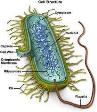
- In 1890, German bacteriologist Robert Koch defined four requirements to prove that a given organism causes a given disease. These requirements, known as Koch's postulates, guide research into the bacterial cause of many diseases, including cancer, and establish a gold standard of evidence consistent with "proof".
-
- Contents
- 1 Known to cause cancer
- 2 Suspected to cause cancer
- 3 History
- 4 Cancer Bacteria as Contaminants
- 5 References
- 6 See also
-
- Known To Cause Cancer
-
-
- Helicobacter pylori colonizes the human stomach and duodenum. In some cases it may cause stomach cancer[1][2] and MALT lymphoma[3]. Animal models have demonstrated Koch's third and fourth postulates for the role of Helicobacter pylori in the causation of stomach cancer.[4]
-
- Suspected Of Causing Cancer
-
-
- Mycoplasma has been implicated in the formation of cancer.[5] Specific reports include:
-
-
- > Mycoplasma can "induce malignant transformation of host mammalian cells" by means of "aberrant expression of oncogenes and tumor repressors"[6].
-
-
- > A "Mycoplasma hyorhinis-encoded protein known as p37..(could) infect humans and may facilitate tumor invasiveness" [7].
-
-
- > A "significantly high existence of Mycoplasma sp. DNA" has been reported "in the tissues of patients with conventional (renal cell carcinoma)" as opposed to normal controls.[8]
-
-
- > "Mycoplasma infection can be detected in many tumor tissues, continuous infection of mycoplasma can lead to transformation of mammalian cells, up-regulating expression of oncogenes, and some biologic changes of tumor cells, suggesting association of Mycoplasma infection with tumorigenesis"[9]
- A number of other bacteria have associations with cancer, although their possible role in carcinogenesis is unclear. Salmonella typhi is associated with gallbladder cancer, Streptococcus bovis is associated with colorectal cancer and Chlamydia pneumoniae with lung cancer.[10]
-
- History
-
-
- In 1890, the Scottish pathologist William Russell reported circumstantial evidence for the bacterial cause of cancer [11]. In 1926, Canadian physician Thomas Glover reported that he could consistently isolate a specific bacterium from the neoplastic tissues of animals and humans.[12] One review summarized Glover's report as follows:
-
-
- "The author reports the isolation of a pleomorphic organism from various types of cancer which can be grown in pure cultures in its several phases. He produced a serum from it which has given remarkable results in a series of 50 reported cases. This is very important, if true. We suppose the Cancer Society will give an opinion later on the reliability of the findings."[13]
-
-
- Glover was asked to continue his work at the Public Health Service (later incorporated into the National Institutes of Health) completing his studies in 1929 and publishing his findings in 1930[14]. He asserted that a vaccine or anti-serum manufactured from his bacterium could be used to treat cancer patients with varying degrees of success [14]. According to historical accounts, scientists from the Public Health Service challenged Glover's claims and asked him to repeat his research to better establish quality control[15]. Glover refused and opted to continue his research independently; not seeking consensus, Glover's claims and results led to controversy and are today, not given serious merit.[16]
-
-
- In 1931, Cornell University pathologist Elise L'Esperance reported the presence of acid-fast, Tuberculosis-like organisms in Hodgkin's disease[17], French physician George Mazet implicated a bacterial association with leukemia and also Hodgkin's disease[18] and a number of other physicians or scientists, including the German von Brehmer[19], the Irish physician W.M. Crofton[20] and EJ Villesquez of France[21] all reported similar cancer bacteria associations. In one case, an Italian scientist named Clara Fonti reportedly inoculated herself in the chest wall with a metastasizing mammary carcinoma and claimed to note neoplastic changes in her own tissues[22].
-
-
- In some cases, claims were made that anti-sera derived from cancer associated bacteria could be used therapeutically. In 1953, for example, the results of an anti-bacterial vaccine trial involving 100 patients said to be diagnosed with different stages and types of cancer was reported by Dr. John E. White at the 6th International Congress of Microbiology in Rome, Italy. White said there were a number of favorable responses, but critics noted that the trial was not blinded, supervised, or monitored by any independent mainstream cancer agency, and the actual severity as well as diagnostic authenticity of the cancers-in-question not verifiable.
-
-
- In 1950, a Newark based physician named Virginia Livingston published a paper claiming that a specific Mycobacterium was associated with neoplasia[23]. Livingston believed that this organism was intermittently acid-fast, highly pleomorphic, and taxonomically related to the leprae and tuberculin bacilli. She, along with several colleagues including microbiologist Eleanor-Alexander Jackson, continued research throughout the 1950's and eventually proposed a name for the bacterium, calling it Progenitor Cryptocides. Livingston also proposed a treatment protocol based on anti-microbial vaccines similar in concept to Glover's which she reported on in a 1965 publication[24].
-
-
- Other investigators were not sure of the exact classification of cancer bacteria, and had claimed to find different taxonomies other than Mycobacteria. For example, I.C. Diller who had reported an increased "number of tumors" occurring in mice "injected with a bacterium" isolated from mice and human cancers, referred to several possible classifications. These included a Cornybacterium "of undetermined species", Staphylococcus epidermidis and an organism related only antigenically to Mycobacteria. She cited the "extreme pleomorphism" and "tendency to stabilize in forms that mimic other bacteria" as reasons for the difficulty in classification[25].
-
-
- Beginning in 1963, the National Cancer Institute (NCI) launched its own preliminary investigation of cancer bacteria, and until 1974, conducted several studies which focused primarily on the culturing of cancer bacteria from animal and/or human cancers in an attempt to establish a guilt-by-association relationship between the two[26][27]. The NCI investigations involved several different species, including Mycoplasma orale, Mycoplasma fermentans, Mycoplasma neurolyticum and Mycoplasma pneumoniae and many of the studies involved examination of leukemic bloods.
-
-
- The NCI studies produced various results. In one study, researchers concluded there was no statistically relevant oncogenic association between certain Mycoplasma species[28]. In another, investigators concluded there was "an association between Mycoplasma and acute leukemia"[29]. Another NCI paper stated the "importance of (Mycoplasma/cancer) association(s)" because "latent mycoplasma were repeatedly isolated from mice under stress with malignant disease"[30].
- As of October 1986, however, the NCI's position was that its bacteria investigation failed to reach a definitive conclusion[31]. NCI and other mainstream investigators---while questioning the actual role of cancer bacteria in oncogenesis---believed that such bacteria were common invaders of cancer tissues, acting more as parasites rather than involved in causality. Mycoplasma are known to be notorious contaminants of laboratory culture and other medium. Throughout the 1980's and up to the present time, the NCI has been actively involved in cancer virus research.
-
-
- In 1974, Livingston reported an association between cancer-related bacteria and human choriogondatropin (HCG), a growth hormone commonly associated with pregnancy[32]. Hernan Acevedo, a Pittsburgh-based scientist, also reported that HCG could be synthesized from cancer-related bacteria[33].
- Livingston believed that HCG was a common denominator linking cancer and bacteria, and alleged that cancer tissues utilize HCG for the purpose of avoiding immunosurveilance, basing her ideas on data suggesting the human embryo uses HCG in the same fashion[34]. In a later critique of Livingston's claims and methods, the NCI argued that "HCG is produced by a variety of bacteria from both cancer patients, and normal tissues".[16]
-
-
- Acevedo reported in additional studies that "the synthesis and expression of hCG, its subunits, and its fragments, is a common biochemical denominator of cancer, providing the scientific basis for studies of its prevention and/or control by active and/or passive immunization against these sialoglycoproteins"[35].
- In 1990, the NCI published a review of Livingston's theories and said that her methods of classifying the cancer bacterium she referred to as "Progenitor cryptocides" had contained "some remarkable errors".[16] Independent analysis of this organism using DNA-DNA hybridization technology revealed its actual classification as Staphylococcus epidermidis, not a Mycobacterium as Livingston had stated.
-
-
- According to Dr. Alva Johnson, a professor at the Department of Microbiology and Immunology at the Eastern Virginia Medical School who had independently reviewed the NCI critique, Livingston had not preserved earlier samples of her cultures taken from the blood and tissues of cancer patients in which she claimed existed Mycobacteria and instead, routinely screened cancer patients' urine samples for HCG-positive bacteria assuming these were positive for Mycobacteria[36]. Johnson noted that Livingston's assumptions reflected "sloppy" science, and adversely affected the protocols needed to build consensus among medical peers[37].
- Livingston died in 1990 and five years later, Dr. Shyh-Ching Lo, a researcher with the Armed Forces Institute of Pathology in Washington, D.C., published a paper titled "Mycoplasmas and oncogenesis: persistent infection and multistage malignant transformation"[38]. According to Lo, Mycoplasma fermentans and Mycoplasma penetrans induced malignant cell transformation in cultured mouse embryo cells, C3H/10T1/2 (C3H) after 6 serial passages lasting 1 wk per passage. He further wrote that up until the 11th passage, "malignant changes were reversible if mycoplasmas were eradicated by antibiotic treatment", but at 18 passages, "irreversible..transformation" occurred.
-
-
- Subsequent to Lo's paper and as of 2007, there were a minimum of 14 published papers in the peer reviewed literature claiming to document an association between various species of Mycoplasma and various animal and human cancers.
-
- Cancer Bacteria As Contaminants
-
-
- Scientists have long suspected that cancer bacteria may be contaminants which infiltrate tissue cell cultures, or invade cancer tissues after the disease has started, as opposed to being a direct cause of cancer. Research is now under way in an effort to clarify this relationship.
-
-
- In a recent study, investigators examined deep tissue specimens taken from patients with Oral Squamous Cell Carcinoma. Using rRNA gene sequencing, and addressing the issue of contamination with sterilizing immersion techniques, the investigators noted a "diversity of bacterial taxa...including several putatively novel species." Some of the bacterial species were "isolated only from either the tumorous or nontumorous tissue" indicating "a degree of restriction." The investigators said that "Successful surface decontamination of the specimens indicates that the bacteria detected were from within the tissue".
-
-
- Rather than finding one taxonomy, the researchers noted a "diversity of bacterial groups" and concluded that the "significance of these bacteria within the tumor warrants further study"[39].
-
- References
-
-
- 1 ^ Egi, Y; Ito M, Tanaka S et al. (May 2007). "Role of Helicobacter pylori infection and chronic inflammation in gastric cancer in the cardia". Japanese Journal of Clinical Oncology 37 (5): 365-369.
- 2 ^ Peter S, Beglinger C (2007). "Helicobacter pylori and gastric cancer: the causal relationship". Digestion 75 (1): 25-35. DOI:10.1159/000101564. PMID 17429205.
- 3 ^ Morgner, A; Bayerdörffer E, Neubauer A, Stolte M (Mar 2000). "Gastric MALT lymphoma and its relationship to Helicobacter pylori infection: management and pathogenesis of the disease". Microscopy research and technique 48 (6): 349-356.
- 4 ^ Watanabe, T; Tada M, Nagai H et al. (Sep 1998). "Helicobacter pylori infection induces gastric cancer in mongolian gerbils". Gastroenterology 115 (3): 642-648.
- 5 ^ Huang, S; Li JY, Wu J et al. (Apr 2001). "Mycoplasma infections and different human carcinomas". World Journal of Gastroenterology 7 (2): 266-269.
- 6 ^ Zhang S, et al.(2006). "Alteration of gene expression profiles during mycoplasma-induced malignant cell transformation." BMC Cancer, May, 4;6:116.
- 7 ^ Ketcham et al. (2005). "p-37 Induces tumor invasiveness", Mol Cancer Ther, Jul;4(7):1031.
- 8 ^ Pehlivan S, et al. (2005). "Can mycoplasma-mediated oncogenesis be responsible for formation of conventional renal cell carcinoma?" Urology, Feb;65(2):411-4.
- 9 ^ Ning JY, Shou CC. (2004). "Mycoplasma infection and cancer". Ai Zheng, May;23(5):602-4.
- 10 ^ Mager, DL (Mar 2006). "Bacteria and cancer: cause, coincidence or cure? A review". Journal of Translational Medicine 4 (14). DOI:10.1186/1479-5876-4-14.
- 11 ^ Russell, W. (1890). "An address on a characteristic organism of cancer". Brit Med J, 2:1356-1360.
- 12 ^ Glover, TJ (1926). "Progress in Cancer Research". Canada Lancet and Practitioner 67: 5.
- 13 ^ Raymond S. Patterson. (1926). "A selected public health bibliography with annotations" American Journal of Public Health 16(12):1242 free full text
- 14 ^ a b Glover, TJ (1930). "The bacteriology of cancer". Canada Lancet and Practitioner 74: 92-111.
- 15 ^ Glover, T.J. and Engle, J.L. (1938). Studies in Malignancy. New York: Murdock Foundation.
- 16 ^ a b c (1990) "Unproven methods of cancer management: Livingston-Wheeler therapy". CA: A Cancer Journal for Clinicians 40: 103-108.
- 17 ^ L'Esperance E.(1931). "Studies in Hodgkin's disease". Annal Surg, 93:162-8.
- 18 ^ Mazet, G. (June/August 1941). "Etude bacteriolgigue sur la maladie d'Hodgkin". Montpellier Medicine
- 19 ^ von Brehmer, W. "Siphonosopra polymorpha n. sp.: ein neuer microorganismus des blutes, seine beziehung zur tumorgenese". Med Welt, 8:1178-1185.
- 20 ^ Crofton, W.M. (1936). "The True Nature of Viruses." Staples Press Ltd, London, England.
- 21 ^ Villesquez, E.J. (1955). "Le Parasitisme Latent des Cellules du Sang chez l' Homme, en Particulier dans le Sang des Cancreeux." Librarie Maloine, Paris, France.
- 22 ^ Fonti, C.J. (1958). "Eziopatogenese del Cancro". Amadeo Nicola.& c. Milan, Italy.
- 23 ^ Wuerthele-Caspe, V et al. (1950). "Cultural properties and pathogenicity of certain microorganisms obtained from various proliferative and neoplastic diseases." Am J Med Sci, 220:638-648.
- 24 ^ Livingston VW, Alexander-Jackson E. (1965). "An experimental biologic approach to the treatment of neoplastic disease". J Am Med Wom Assoc, 20:858-866.
- 25 ^ Diller, I.C. (1974). "Tumor Incidence in ICR/Albino and C57/B16JNIcr Male Mice Injected With Organisms Cultured From Mouse Malignant Tissues". Growth, 38:507-517.
- 26 ^ Personal communication from: Carol Case, Chief, Public Inquiries section, Office of Cancer Communications, National Cancer Institute,(October, 1986).
- 27 ^ Source: Journal of the National Cancer Institute. (1963-1974).
- 28 ^ Ebbesen, P. and Lind, K. (1969). "Lack of Evidence for Oncogenic or Amyloid Inducing Qualities of Mycoplasma Neurolyticum Inoculated Into Balb/C Mice". Acta Path Micorbiol Scand, 76:594-600.
- 29 ^ Barile, M.F., et al. (1965). "Isolation of Mycoplasma orale From Leukemic Bone Marrow and Blood by Direct Culture". JNCI, 36:155-159.
- 30 ^ Tully, J. (1967). "Mycoplasma In Leukemic and Nonleukemic Mice". Ann NY Acad Sci, July 28;143(1):345-352.
- 31 ^ Case, ibid.
- 32 ^ Livingston, V.W. (1974). "Some cultural, immunological, and biochemical properties of Progenitor cryptocides." Trans NY Acad Sci, 36:569-582.
- 33 ^ Acevedo, H.F. et al. (1978). "Immunohistochemical localization of a choriogonadotropin-like protein in bacteria isolated from cancer patients". Cancer, 41:1217-1229.
- 34 ^ White, A and Loke Y. (1978). "Increased Sialylation of Surface Glycopeptides of Human Trophoblasts Compared with Fetal Cells from the Same Conceptus". J Exp Med, 148:1087-92.
- 35 ^ Acevedo H.F., et al. (1995). "Human chorionic gonadotropin-beta subunit gene expression in cultured human fetal and cancer cells of different types and origins," Cancer, 76:1476-1475, Oct 15.
- 36 ^ Personal communication from: Dr. Alva Johnson, Dept of Microbiology and Immunology, Eastern Virginia Medical School, May 27, 1992.
- 37 ^ ibid.
- 38 ^ Lo SC, et al. (1995). "Mycoplasmas and oncogenesis: persistent infection and multistage malignant transformation". Proc Natl Acad Sci, 92(22):10197-10201.
- 39 ^ Hooper, S.J. et al. (2006). "Viable Bacteria Present within Oral Squamous Cell Carcinoma Tissue". Journal of Clinical Microbiology, 44;5:17191725
-
-
- Alan Cantwell M.D.
-
- alancantwell@sbcglobal.net
- http://www.ariesrisingpress.com
-
- author of, The Cancer Microbe: The Hidden Killer in Cancer, AIDS, and other Immunologic Diseases, and
- Four Women Against Cancer: Bacteria, Cancer and the Origin of Life
|
|
18 Foods That Make Your Skin Glow
Posted Wed, Mar 19, 2008, 1:20 pm PDT
Top skin creams average about $400 an ounce (and you thought gas was expensive!), yet most offer little proof that they do half of what they promise. Want to save a bundle and improve your skin? Load your shopping cart with nutrients that research has shown to have skin-hydrating, sun-protecting, and even wrinkle-preventing powers, says Manhattan dermatologist Amy Wechsler, MD. Here’s her grocery list of best foods for your skin.
Firm and Bright
You’re probably up to your eyebrows (Botoxed or not) with hearing “eat more fruits and vegetables.” But if you have yet to take that advice to heart, maybe knowing that they prevent wrinkles will do the trick. The colorful pigments that produce bright orange and red also refill antioxidant levels in your skin.
The skin doc’s 3 top picks: SWEET POTATOES, TOMATOES, CANTALOUPE
What they do: Replenish your skin’s supply of antioxidants, so they're ready to scarf up free radicals whenever they make an appearance. Free rads are highly reactive oxygen molecules that damage cells and contribute to just about everything that can go wrong with skin, from dryness to crinkles.
Fresh and Juicy
Your body can’t store much wrinkle-fighting vitamin C, so you need to keep your supplies stocked. The easiest, simplest way: Have some citrus every day.
The skin doc’s 4 top picks: ORANGES, LEMONS, LIMES, GRAPEFRUIT
Actually, ounce for ounce, oranges are the top citrus C source but you can only eat so many, right? For variety, make lemonade, squeeze limes on melon, add grapefruit to salad, and instead of drinking soda, fizz up OJ with sparkling water. It all adds up.
What they do: Keep skin’s vitamin C levels high. While C’s a nifty antioxidant, that’s not the key reason it’s here. It helps keeps collagen -- the supportive protein fibers that stop skin from sagging -- strong and resilient. (Flimsy collagen means lines and wrinkles.) Since collagen breakdown really picks up in your mid-30s, eat citrus early and often to head off aging.
Smoothing and Soothing
There’s a particularly potent antioxidant known as EGCG that does all kinds of good things for skin. The best place to find it? True teas: black, green, or white (not herbal). Brew a teapot full every morning, so that sipping four to six cups throughout the day is a no-brainer.
The skin doc’s #1 pick: GREEN TEA
While all true teas contain EGCG (epigallocatechin-3-gallate), the various types of green tea have the most. Wechsler’s personal favorite is green tea ). “The roasting process that turns this green tea a brownish color also lowers its caffeine content,” she says -- handy if you’re caffeine sensitive or it’s one of those days when you do not need another stimulant.
What it does: Gives your skin a healthy dose of EGCG, which is a great multi-tasker. EGCG puts a damper on inflammatory chemicals involved in acne and sun-related skin aging; it also helps prevent skin cancer; and it has a lion-tamer effect on tumor cells. What’s more, green tea contains L-theanine, a de-tensing amino acid -- and anything you can do to staunch the flow of the stress hormone cortisol helps keep collagen fibers intact.
Green and Leafy
Certain dark leafy greens, whether they’re fresh, frozen, raw, or steamed, really deliver on vitamin A, one of the most skin-essential vitamins going.
The skin doc’s 3 top picks: SPINACH, TURNIP GREENS, BROCCOLI
What they do: Deliver a hefty supply of vitamin A, which supports skin cell turnover, the process that keeps cell growth and development humming along flawlessly. Without enough A, skin becomes dry, tough, and scaly.
Fisherman’s Faves
Several cold-water catches give your skin a double benefit: age-fighting omega-3 fatty acids plus the restorative powers of protein.
The skin doc’s 7 top picks: SALMON, TROUT, TUNA, ATLANTIC MACKEREL, SARDINES, PACIFIC HERRING, MOST SHELLFISH
Just don’t, uh, go overboard. As good as omega-3s are for skin (and the rest of you, too), worries about the amount of mercury in many fish mean it’s smart to limit seafood to two meals a week. That’s a must for women who are or might become pregnant or are nursing, and for young children, too.
What they do: Omega-3s fight inflammation, now considered one of the top skin-agers, and they also help protect against sunburn, enhancing the effects of your SPF sunscreen. Protein is required to build and repair skin cells and to make enzymes and hormones that help keep it glowing.
Fill your weekly grocery cart with all of the above foods and you won’t just look younger, you’ll be younger. Eating at least 1 serving of fish a week and getting the right amount of antioxidants through diet or supplements lower your biological age. In fact, the antioxidants alone can make your RealAge up to 6 years younger.
|
|
For a short-term problem,
tissue salts can help restore order. Take 4 tablets under
the tongue three times daily for two to three weeks.
Kali phos helps
nervous depression from grief and worry. It is the principal
tissue salt for the nerves and should be used alternately
with other remedies.
Nat mur is indicated
for depressed spirits and feelings of hopelessness. This
sadness is typically accompanied by headaches and
constipation.
Nat sulph relieves
depression following an injury to the head.
Calc phos helps with
wandering thoughts and poor concentration.
Next Topic: |
Nanotech Medical Center Lahore Pakistan Leading Facility in
Disease Prevention , and treatment.
http://www.cidpusa.org/FIBROMYALGIA1.html
 |
|